You are here
In the brain, connections rule the roost!
In an article published in early November in Science,1 you proposed a new model for brain functioning. What was the notion that predominated so far?
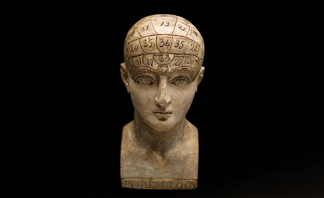
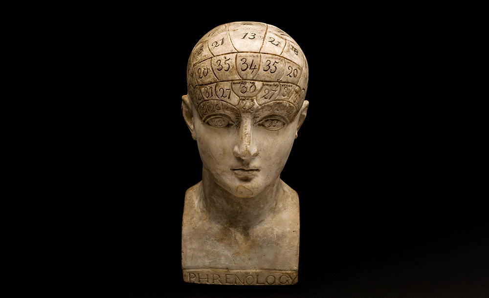
Michel Thiebaut de Schotten:2 We are trying to go beyond the ‘locational’ model that attributes each cognitive function, such as language, memory or reasoning, to a specific area of the brain. Dating from the early 19th century and derived from phrenology – a pseudo-scientific theory according to which the bumps on the skull reflect personality traits (the most famous example being the ‘maths bump’) – this concept was initially corroborated by several scientists. In 1861, the French physician Paul Broca thus observed that the brain region involved in language production was situated at the front of the cranium, and gave his name to what has been known since as the ‘Broca area’. However, over the past twenty years, several studies have called this reductionist vision into question.
What does your new model suggest?
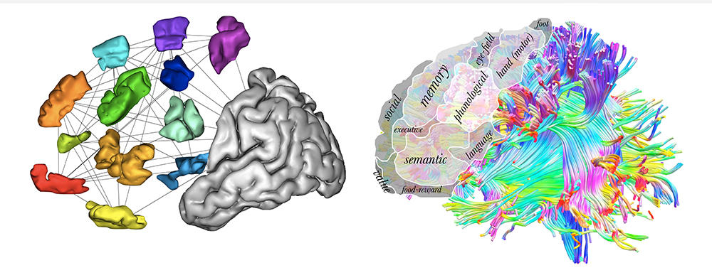
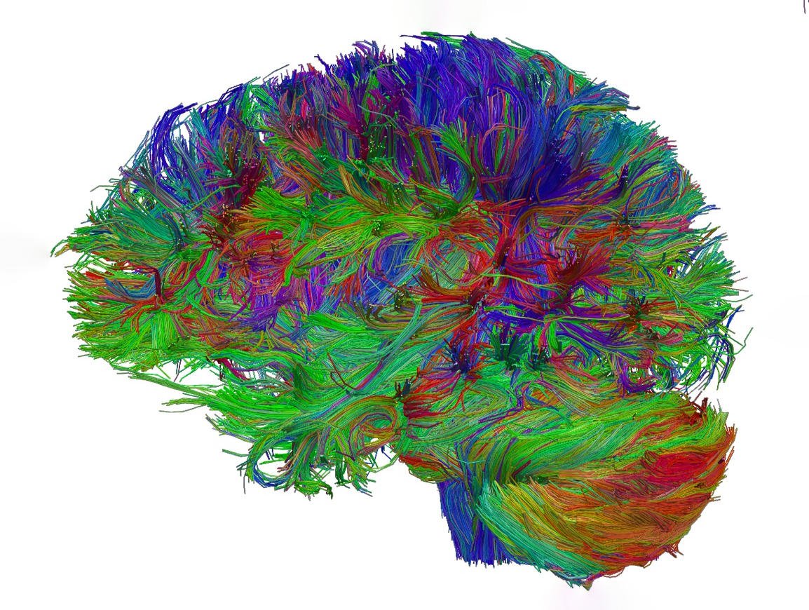
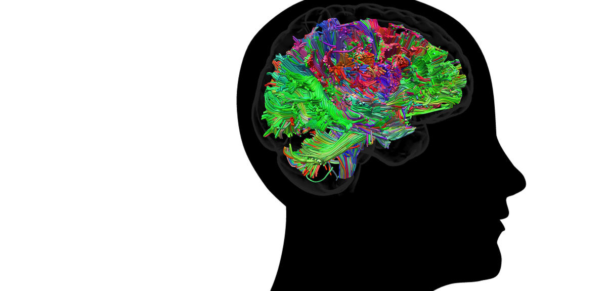
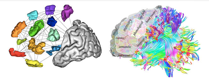
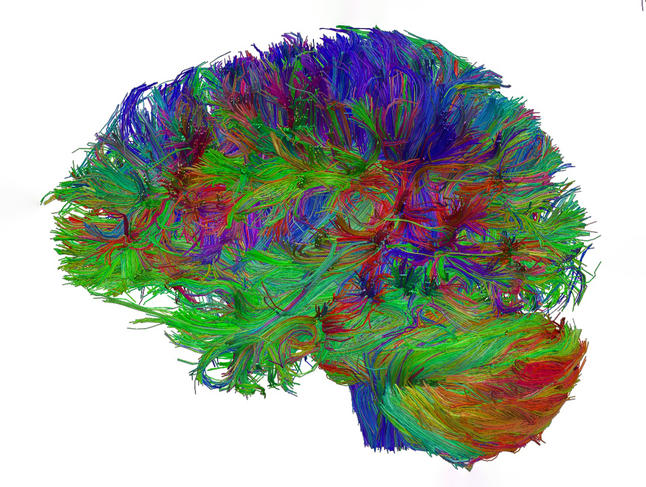
M. T. de S.: Unlike the classic model referred to above, we propose that brain functions emerge not just from areas but from exchanges between these areas, thus placing connectivity at the heart of brain functioning. Located under the cerebral cortex the external layer of the brain, or “grey matter”; editor’s note, and forming the white matter, these connections are established via neuronal extensions, or axons, which are grouped in bundles that connect the areas of the brain.
How does this connectionist model overturn previous concepts?
M. T. de S.: Under this model, brain connections are no longer considered as simple channels for the transfer of signals between different cerebral areas. On the contrary, they appear to act as conductors of the orchestra, capable of amplifying or diminishing brain signals and favouring ‘dialogue’ between several areas by interconnecting them at a given time for a given duration. In doing so, they enable the emergence of functions that could not be carried out in isolation by particular brain regions, as these may be activated by a wide variety of tasks. The brain thus appears to be a much more dynamic ensemble from which many additional capabilities can emerge! In short, its integrated functioning using connections means that, as described by the Greek philosopher Aristotle, ‘the whole is greater than the sum of its parts’.
When did this new model start to take shape?
M. T. de S.: In the early 2000s, thanks to a series of studies which showed that functions are dependent on circuits and not just on brain regions. For example, an American research in the ferret, published in 2000,3 found that disconnecting the axons that innervate the visual cortical areas responsible for processing visual information from the eye; editor’s note and reconnecting them to the auditory brain areas which process sound caused a change to the organisation of the cells forming the latter: they then acquired a “cytoarchitecture” similar to that of the visual zones, which enabled the animals to see correctly. Hence the conclusion that it is the brain connections that are in control of the different brain regions.
When did you start to agree with the connectionist model?
M. T. de S.: As far back as 2004, at a time when it was still little accepted. I started to understand the major contribution of cerebral connections to brain functioning following studies4 on two patients operated on by the neurosurgeon Hugues. We discovered ‘by chance’ that unilateral spatial neglect syndrome (or hemineglect), where the patient cannot detect half of the visual field, resulted not from a lesion in the cortical area but the disconnection of cerebral networks between the front and back of the brain. Since then, we have published several other studies based on these initial findings. For example, in a research paper published in 2015,5 we showed that the dysfunction of cerebral networks was the common cause for the behavioural and cognitive disorders reported in three historic neurology patients.6
How do you study brain connectivity?
M. T. de S.: Mainly thanks to two powerful brain imaging techniques that enable us to see the brain as it functions without opening the skull: functional magnetic resonance imaging (fMRI) provides information on the neuronal networks at work during a task; while diffusion MRI (dMRI) informs on the structure of connections (trajectory, diameter, etc.). That said, access to these technologies is difficult because they are expensive and only a handful are available in France.This is why, in recent years, we have developed two applications – bcbtoolkit and Funtionnectome – that can detect and indirectly study potential brain disconnections when a patient has undergone an MRI examination once in their life. Available free of charge online,7 these tools developed by my team are based on a series of algorithms and on an atlas that maps the principal brain connections in humans.
Have there been interesting findings regarding brain connectivity in healthy individuals?
M. T. de S.: It is now well established that the architecture of neural networks varies from one person to another, somewhat like genetic information, where there is only a 0.1% difference between two individuals. This ‘neurovariability’ determines who we are, because variations in connectivity are associated with alterations in performance regarding certain cognitive functions (memory, language, fine motor skills, etc.). But one important point is that our brain connectivity can change considerably throughout life, in particular as a function of learning. This can cause the appearance or disappearance of certain connections, thanks to brain plasticity processes.
Could the connectionist model revolutionise research on the brain?
M. T. de S.: Absolutely! First of all, it should help us to clarify brain functioning and individual differences in terms of cognitive performance. Studying brain connectivity may also improve our knowledge of the evolution of the human encephalon: by comparing the profiles of connections in our brain with those of various species of similar primates (macaque, chimpanzee, etc.) it is possible to go back in time and model the brain connectivity of our common ancestors.
How important is your model from a clinical point of view?
M. T. de S.: It may improve our management of neurological and psychiatric disorders, in several ways. For example, it could enable physicians to explore the brain connections of a patient, and more quickly acknowledge – in case broken or poorly connected links are detected – that the patient is suffering from a real disorder. Furthermore, this new model may predict the evolution of the after-effects of a stroke (cerebrovascular accident, or CVA). Indeed, we know that the brain connectivity of an individual impacts their ability to recover more or less rapidly from such an event. In this case, my team has developed a tool which, based on a map of the disconnections in a patient, can estimate the probability that they might walk, talk, or be able to move a paralysed arm again, a year after the stroke. This interactive web app, called ‘Disconnectome Symptoms Discoverer’ is also available online.8 All these advances bode well for more personalised care and optimisation of the monitoring and management of patients.
- 1. “The emergent properties of the connected brain”, M. Thiebaut de Schotten and S. Forkel, Science, 4 November 2022. doi: 10.1126/science.abq2591. Epub 3 November 2022.
- 2. CNRS scientist at the Institute of Neurodegenerative diseases (CNRS / Université de Bordeaux) within the Neurofunctional imaging group, and a member of the Brain Connectivity and Behaviour Laboratory at Université Paris Sorbonne.
- 3. “Induction of visual orientation modules in auditory cortex”, J. Sharma, A. Angelucci, and M. Sur, Nature, 20 April 2000. doi: 10.1038/35009043. “Visual behaviour mediated by retinal projections directed to the auditory pathway”, L. von Melchner, S. L. Pallas and M. Sur, Nature, 20 April 2000. doi: 10.1038/35009102.
- 4. “Direct evidence for a parietal-frontal pathway subserving spatial awareness in humans”, M. Thiebaut de Schotten et al., Science, 30 September 2005. doi: 10.1126/science.1116251.
- 5. “From Phineas Gage and Monsieur Leborgne to H.M.: Revisiting Disconnection Syndromes”, M. Thiebaut de Schotten et al., Cereb. Cortex, December 2015. doi: 10.1093/cercor/bhv173. Epub 2015 Aug 12.
- 6. In the 19th century, after a serious cranial injury, the American Phineas Gage underwent a profound personality change; also in the 19th century, the Frenchman Louis Victor Leborgne developed severe language disorders following an epileptic fit, and finally, the American Henry Gustav Molaison, who died in 2008, lost his ability to remember new information following a neurosurgical procedure.
- 7. On the “Brain Connectivity and Behaviour Laboratory” website, at: www.bcblab.com.
- 8. ibid.
Explore more
Author
A freelance science journalist for ten years, Kheira Bettayeb specializes in the fields of medicine, biology, neuroscience, zoology, astronomy, physics and technology. She writes primarily for prominent national (France) magazines.


