

You are here
The many tricks of roaming cells

The phenomenon is fascinating. When observing a fibroblast cell from the skin or cartilage in culture on a Petri dish under a microscope for several minutes, one can actually see it crawl! And it does that on its own, without the intervention of external agents but thanks to several coordinated cyclical processes, repeated over and over.
First of all, the cell projects a broad, flat extension that binds to its substrate, forming what is called a lamellipodium. The cell contracts, while at the same time detaching its tail. This cycle of protrusion, adhesion and contraction then repeats itself, and the cell moves forward on its substrate, rather like a snail.
A fundamental…
This motility process, which takes place when cells are intertwined in tissues, enables them to move at a speed of 10 to 15 micrometres (thousandths of millimetres) per minute. As a comparison, spermatozoa, which use quite another type of motility (swimming, thanks to a sort of tail called a flagellum) can move at 3 mm per minute, or between 100 and 200 times more rapidly.
The fact remains that “cell motility within tissues is a fundamental process. It is essential to the formation and maintenance of the human body and of other multicellular (made up of several cells) organisms”, explains Matthieu Piel, a cell biologist in the Cell Biology and Cancer laboratory1, at Institut Curie in Paris. “Clarifying its mechanisms could provide new perspectives in medicine, notably regarding the treatment of cancer.”
…and lifelong process
Cell motility occurs from the first moments of life, during embryonic development – the process that starts with fertilisation and continues until birth. “At that time, embryonic cells move actively over distances of several centimetres in order to reach specific sites which correspond to those of future organs, so as to form different tissues and organs,” the biologist explains.
Thereafter, there seems to be a degree of stability, although it is not apparent, as cell motility persists throughout life. It is fundamental in particular to the regeneration of tissues such as the intestinal mucosa (which coats the inner surface of the intestine). “In this case, cell motility is essential to allow new cells produced at the base of this tissue to travel to its surface. A continuous process, this directional migration of epithelial cells guarantees the integrity and functionality of the intestinal mucosa,” explains Danijela Vignjevic, who is studying this phenomenon in the Cell Biology and Cancer laboratory.



Motility is also a key component in skin healing. Indeed, this requires the movement of several cell types: immune cells which are transported by the blood to enter the damaged tissue in order to eliminate debris and stimulate repair; fibroblasts that move towards the wound site in order to proliferate and contribute to producing a new extracellular matrix (network of macromolecules surrounding cells in the tissues); or keratinocytes, which move from the edges of the wound to create a new and complete epidermis.
Several types of motility
There are several modes of cell motility, depending on the type of cells and their environment. That described above is also called “mesenchymal motility” as it is adopted by mesenchymal cells (the mesenchyme is an embryonic connective tissue).
Discovered in the 1970s by the British cell biologist and embryologist Michael Abercrombie (1912-1979), mesenchymal motility is now well understood at a molecular level. It has notably been established that it is the elongation (or ‘polymerisation’) of actin microfilaments that initially allows projection of an extension of the cell membrane. Actin molecules are a major component of the cytoskeleton, the network of proteins responsible for maintaining cell shape.
It is also known that adhesion protein complexes – or integrins – then allow adhesion of the lamellipodium to its cellular environment. Situated at the ends of actin fibres in the cytoskeleton, these large molecular structures enable the active anchorage of this cell to its substrate and transmit a mechanical force that propels it forwards. Indeed, at the same time, the adhesion structures at the rear of the cell disassemble. The latter portion of the cell then detaches itself and the cell contracts, which allows it to move forwards.
Finally, this contraction draws strength from another molecule that acts as a molecular motor: myosin. This molecule binds to actin microfilaments and uses ATP (the energy molecule in cells) to ensure they slide past myosin filaments, causing retraction of the cell membrane.


Parasite-like
A second type of cell motility is equally fascinating: amoeboid movement. “Although this process has been discovered more recently (a few decades ago), it nevertheless seems to be the most widespread and ancestral mode of migration for cells that ‘crawl’”, underlines Matthieu Piel. “It is of considerable interest to scientists as it is the type of motility used by immune cells and the most aggressive tumour cells: amoeboid cancer cells.”
As its name suggests, the amoeboid mode concerns cells whose movements recall those of amoeba, the highly mobile single-cell parasites that are found in soiled water. In the same way as these micro-organisms, the cells adopt this crawling motility by deforming their plasma membrane and projecting extensions that resemble “cellular feet”, or pseudopods (from the Greek “pseudo” for false and “podos” for foot); these structures are often bulbous “blebs”) or filiform, while the lamellipodia of mesenchymal motility are flat and broad.
“In the case of tumour cells, we do not use the term pseudopods but ‘invadopodia’, to emphasise their ability to invade other organs,” notes Philippe Chavrier, biologist in the Cell Biology and Cancer laboratory, and co-author of a recent article2 reviewing current knowledge of these structures.


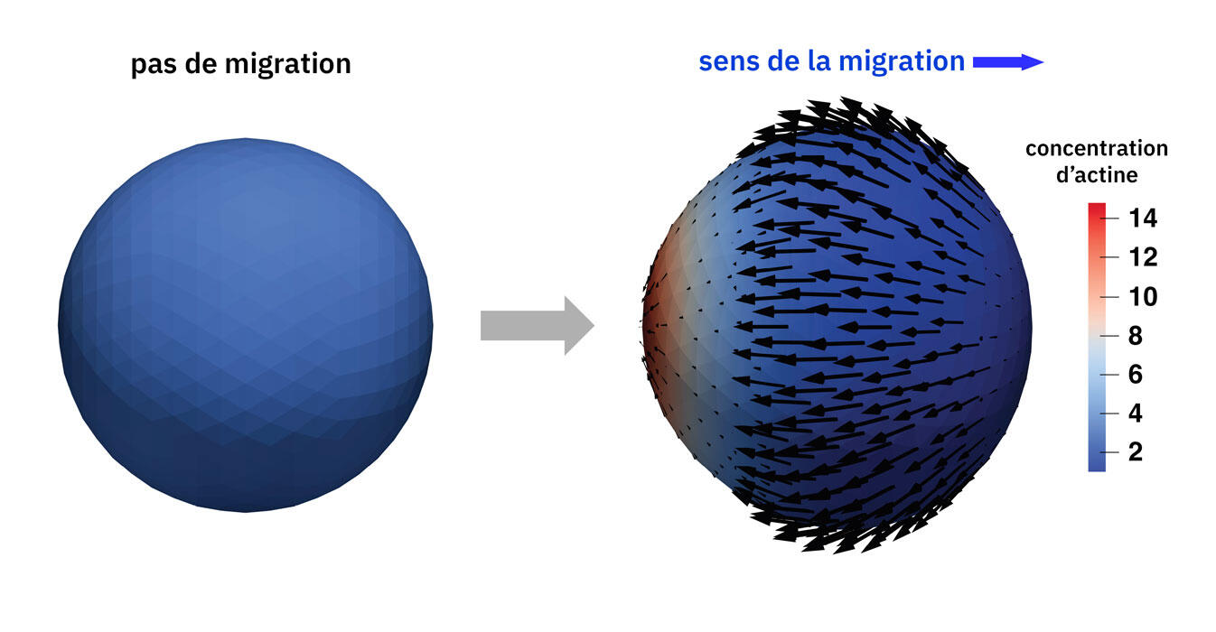
Using digital simulations3, Alexander Farutin and his colleagues in the Interdisciplinary Laboratory of Physics (LIPhy)4, near Grenoble (southeastern France), have been able to show that the addition of actin molecules at the growing end of filaments, accompanied by their dissociation at the other end, was sufficient to induce cell movement, all without the intervention of myosin. “This discovery questions the classic dogma according to which molecular motors, and particularly myosin, are essential to cell motility,” notes the physicist.
Climbers and swimmers
More recently, in the early 2000s, scientists discovered a third and intriguing mode of motility referred to as “chimney climbing” because of its resemblance to the technique adopted by mountaineers to climb up rocky crevices. “This type of motility enables the cell to move by pushing on its environment, a little like pushing off rock walls in order to climb,” describes Matthieu Piel, who helped shed light on this process5.
Unlike the first two modes, this movement does not require any adhesion. It is mainly adopted by immune cells and mobile amoeboid cancer cells when they are trapped between two substrates: narrow blood microcapillaries, tight spaces between fibres in the extracellular matrix or between the cells of dense tissues, etc.

Swimming
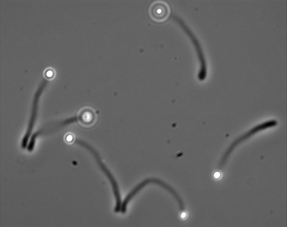
There remains a final type of motility: swimming! Somewhat similar to spermatozoa, but this time propulsion is achieved thanks to molecular paddles and not a flagellum. Furthermore, it takes place within tissues and not in the male and female reproductive tracts.
“Our work6 has shown that immune cells are able to swim thanks to transmembrane proteins, such as integrins, whose intracellular part is linked to the cytoskeleton, while the extracellular part acts like a paddle,” explains Olivier Théodoly from the Adhesion & Inflammation Lab (LAI)7 in Marseille (southeastern France). According to some immunologists, this type of process may explain how immune cells move within the interstitial fluid that fills the spaces between cells inside inflamed tissues.
Fighting metastases
Elucidating the mechanisms of cell motility could help to develop treatments that are able to restrict or prevent the formation and migration of cancer metastases.
In 2022, the team led by Cécile Sykes, a physicist specialised in soft materials (whose shape can be changed) in the (LPENS)8 in Paris, published a “recipe” for the development of a system that might prove of great help in addressing this challenge. This device involving microscopic beads coated with a special (WASP) protein makes it possible to initiate the assembly of actin, and thus enables the reconstitution of actin-based motility9. “Our system might lead to the identification of drugs that are able to block the polymerisation of actin specifically, and thus the movement of cancer cells,” explains the physicist.

Mechanical therapies round the corner?
Alongside medicines, a clearer understanding of cell motility could also open the way to mechanical rather than chemical opportunities. This is the challenge being taken up by teams in the Jean-Alexandre Dieudonné Laboratory (LJAD)10, in Nice (southeastern France), and the LMPS11. “We hope to develop 'theramechanics' (a contraction of mechanical therapies – Ed’s note) such as injectable hydrogels or materials that can modify the mechanical properties (shape, resistance, etc.) of cells, their components or their environment, and thus restrict their migration,” explains Rachele Allena from the LJAD.
She recently proposed a theramechanics model that consisted in modulating the geometric and mechanical properties of the nucleus12. In fact, this rigid and highly voluminous cell component could thus be manipulated to prevent the movement of cells (including cancer cells) within the narrow interstices of tissues. Although still exploratory, this option is highly promising.
See also
Shedding new light on the adventure of humanity
- 1. CNRS / Institut Curie.
- 2. C. Gamblin et P. Chavrier, “Invadosomes – Entre mobilité et invasion, naviguer dans la dualité des fonctions cellulaires”, Med Sci, 2024: https://doi.org/10.1051/medsci/2024080
- 3. W. Schmidt, et al., “Myosin-Independent Amoeboid Cell Motility”, Physical Review Letters, 2025: https://doi.org/10.1103/PhysRevLett.134.158301
- 4. CNRS / Université Grenoble Alpes.
- 5. R. J. Hawkins, et al., “Pushing off the Walls: A Mechanism of Cell Motility in Confinement”, Physical Review Letters, 2009: https://doi.org/10.1103/PhysRevLett.102.058103
- 6. L. Aoun, et al. “Amoeboid Swimming Is Propelled by Molecular Paddling in Lymphocytes”, Biophysical Journal, 2020: https://doi.org/10.1016/j.bpj.2020.07.033
- 7. CNRS / Aix-Marseille Université / INSERM.
- 8. Laboratoire de Physique at École Normale Supérieure (CNRS / ENS-PSL / Sorbonne Université / Université Paris Cité).
- 9. C. Sykes et J. Plastino, “Reconstitution of Actin-Based Motility with Commercially Available Proteins”, Journal of Visualized Experiments, 2022: https://dx.doi.org/10.3791/64261
- 10. CNRS / Université Côte d’Azur.
- 11. Laboratoire de Mécanique Paris-Saclay (CNRS / ENS Paris-Saclay / CentraleSupélec).
- 12. Rachele Allena, “Potential theramechanics for cancer invasion: an in silico study”, Mathematics in Medical and Life Sciences, 2024: https://doi.org/10.1080/29937574.2024.2404052
Explore more





Author
A freelance science journalist for ten years, Kheira Bettayeb specializes in the fields of medicine, biology, neuroscience, zoology, astronomy, physics and technology. She writes primarily for prominent national (France) magazines.