You are here
Three Innovation Start-ups on the Move
OliKrom: chameleon pigments that reveal the invisible
Materials that change color based on impact, pressure, or temperature: OliKrom’s intelligent pigments have created a small revolution in their field.
Imagine that you’re driving along a winter road in a downpour, but you’re confident because photoluminescent pigments have optimized road markings, making even black ice visible. Coatings, paints, and inks that adapt to their environment and use color as an indicator—that is the heart of OliKrom’s technological mission, at the intersection of basic research and its industrial applications.
“Our pigments respond to environmental problematics in fields ranging from aeronautics to building coating and food packaging,” explains Jean-François Létard, co-founder and a former senior researcher at the Institut de Chimie de la Matière Condensée de Bordeaux (ICMCB), where the research behind the start-up began.

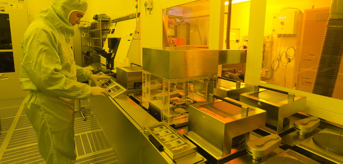
Létard and his team successfully filed for their first international patent in 2005. In 2013, he completed the HEC challenge + for management and entrepreneurship training, and created OliKrom. After securing fundraising from Starquest Capital and BPI France, OliKrom now has 14 employees, as well as a dedicated industrial site. The company has been working with Airbus for nearly 6 years, notably on “pigments that can detect impacts on composite materials through pressure variation, thereby quantifying the damage sustained by airplane parts during production or transportation.” OliKrom is also developing coatings that can signal the presence of a gas, solvent, or light. For OliKrom’s chameleon pigments, the adventure has just begun.
Sensome: a miniature sensor that is a valuable tool for stroke treatment
Sensome has developed a technology to help physicians make decisions during the crucial hours after a stroke. The start-up will begin clinical testing soon.
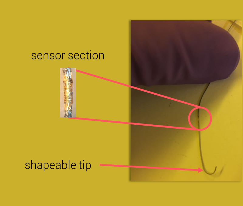
Each year, 1.5 million people in the EU and the US suffer an ischemic stroke. A novel intervention in the ensuing hours can remove the blood clot from the obstructed vessel. The optimal removal tool, however, depends on the type of clot blocking the artery. Hence Sensome, a spin-off from the Laboratoire d’hydrodynamique de l’Ecole polytechnique (LadHyX),1 proposes an ultra-miniaturized AI-equipped sensor, which placed at the tip of a neurovascular guidewire, can precisely identify the nature of the clot. This smart guidewire, called Clotild™, provides crucial insights for the removal procedure, enabling the interventionalist to adapt the tool to each patient.

Sensome co-founder Franz Bozsak began his research with stents, trying to devise ways of equipping them with sensors. “You needed a sensor that is tiny but also sensitive enough to relay reliable information.” Bozsak focused on impedance sensors used to follow cell movement, which he adapted to read tissue properties. “Depending on the impedance measurement, we can distinguish white clots, which are rich in fibrin and do not break easily, from red clots, which are rich in red blood cells and brittle.” Bozsak simultaneously completed a joint Stanford University-École polytechnique program to learn more about the business aspects of his bold adventure, and used funding from multiple competition prizes to found his start-up. He and his team of engineers worked hard to miniaturize this sensor, understand which signals convey information, and develop artificial intelligence algorithms for the analysis. The start-up has “six patent families, some of which co-owned by the CNRS,” which also contributed to the company’s capital.
Damae Medical working to diagnose skin tumors without a biopsy
The start-up’s rapid and non-invasive technology is now in the pre-industrialization phase.
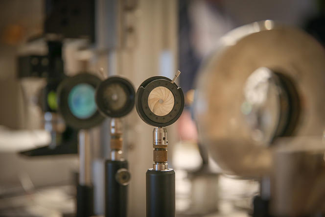
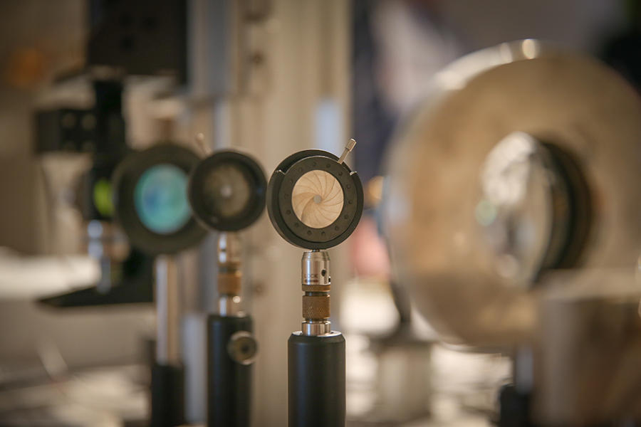
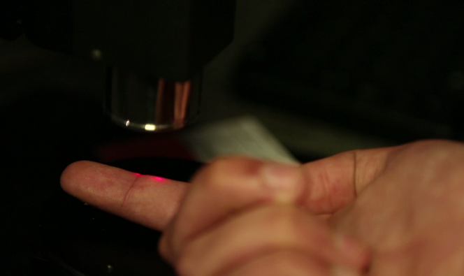
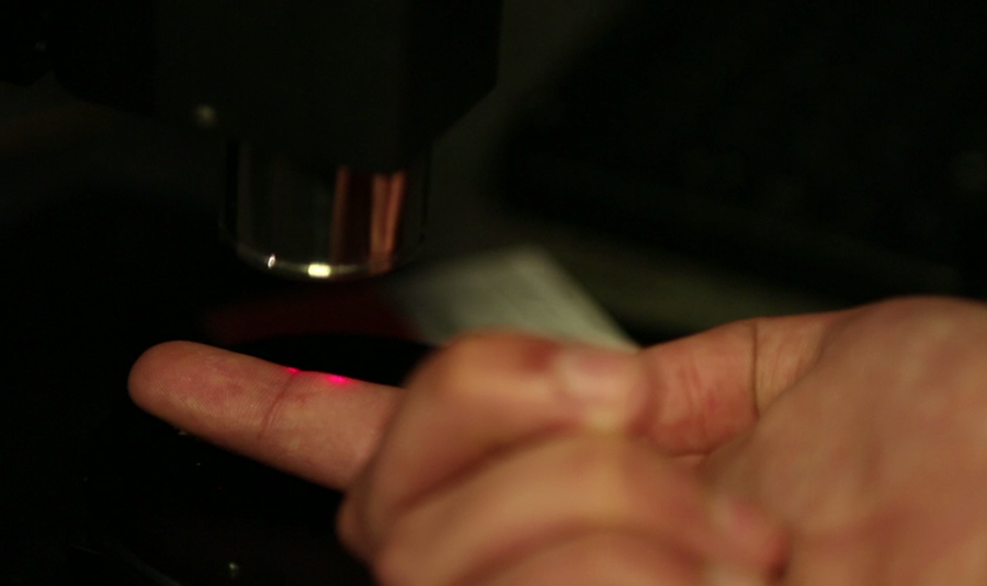
Diagnosing skin cancer requires a biopsy, which is to say analyzing a patient’s tissue sample with a microscope. Damae Medical, which grew out of research conducted at the Laboratoire Charles-Fabry,2 is aiming to revolutionize this exam. The technology developed by Damae Medical will provide dermatologists with an in vivo imaging modality that produces in-depth images of the tissue similar to histology without the need for skin tissue excision and processing. Co-founder and Chief Scientific Officer Pr. Arnaud Dubois focused on a single question: how to image biological tissue mass on the cellular scale of a few microns. Such tissue actually acts as a diffusive environment on light, similar to frosted glass, as rays do not propagate in straight lines and quickly lose all “memory” of their origin, thereby blurring images.
A partial answer came in the form of optical coherence tomography (OCT), which uses the interference of two light beams—one of which is reflected by the tissue being analyzed—to reconstruct an image layer-by-layer based solely on the photons that underwent no deviation. Still, OCT cannot always provide micron-level detail, hence the researcher’s idea to pair it with the technique of reflectance confocal microscopy, which images a tissue point-by-point by eliminating parasitic light through a specially placed slot. “Combining the two techniques allows for dual filtering, once by interference and once spatially,” Dubois points out. Most importantly, it provides clean micron-scale images up to a millimeter in depth.
Dubois turned to the “Entrepreneur innovation network” of his home institution l’Institut d’Optique Graduate School to create the start-up in 2014. A series of innovation awards—including one from the MIT Technology Review—eventually culminated in clinical proof-of-concept and successful testing directly on patients.
Explore more
Author
Arby Gharibian is a writer, translator, and independent researcher in social sciences, art, and literature .


