You are here
Putting our organs on chips
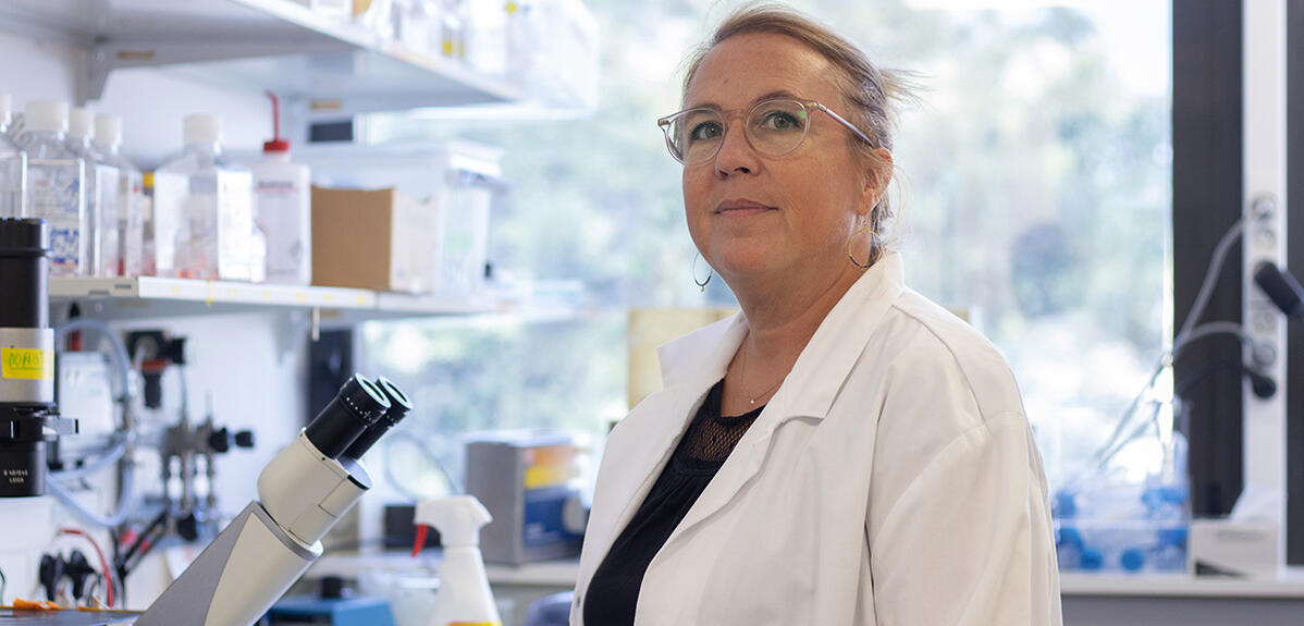
When talking to Stéphanie Descroix1, CNRS research professor and team leader at Institut Curie in Paris, what strikes you immediately is her positive attitude. About her workplace: “wonderful, the best one to carry out my research”, she says. About her work: “fantastic” and “extremely satisfying”. Her career: “I have been very lucky!” And her colleagues, many of whom are “remarkable”. “She creates such a good atmosphere in her group that it is difficult to leave,” observes Charlotte Bouquerel, who has been working with her for four years in the context of her doctoral thesis.
Yet if the scientist stands out, it is also because of her research... at the cutting edge of technology. Her Macromolecules and Microsystems in Biology and Medicine (MMBM) team is one of the global leaders in a recent field that promises to revolutionise our understanding of physiology and human pathologies and their management: organs on chips.
“Organs on chips are new technologies designed to reproduce some of the cellular, biochemical, physical and physiological characteristics of human organs and tissues, such as their three-dimensional structure, physicochemical environment (oxygen content, acidity, etc.) or their functions”, Descroix explains. These systems are produced thanks to microfluidics, a technology born some thirty years ago and seeing exponential growth today.
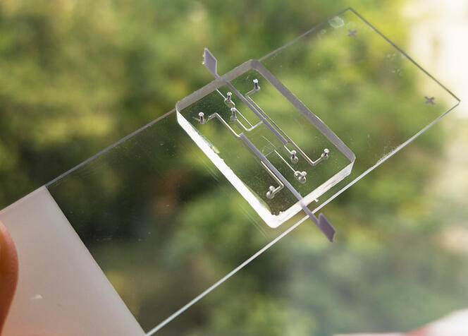
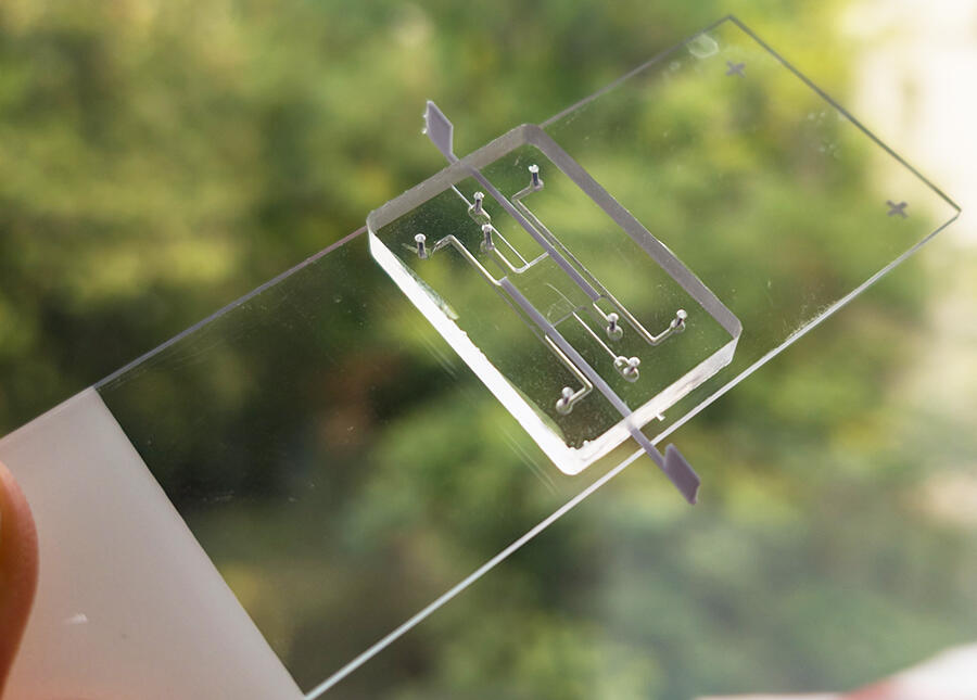
At the crossroads between biology, physics, chemistry and engineering, microfluidics enables the manufacture of miniature devices on glass, silicone or plastic chips. Although very small (a few centimetres square), these platforms contain a series of engraved or moulded microchannels that are interconnected so that they can perform a given function, such as mixing components or controlling the biochemical environment.
The art of doing better with less
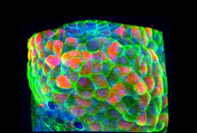
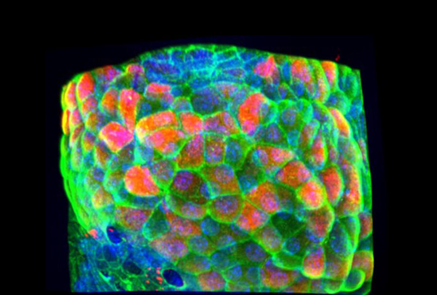
In practice, “organs on a chip are obtained using cells and molecules from the extracellular matrix, the ‘cement’ that binds cells together in a particular tissue. This assembly is injected into a microfluidic chip where it self-organises to acquire a three-dimensional structure that may be similar to that of a real organ”, the biologist details.
Organs on a chip have huge advantages! Firstly, microfluidics enables the control of different biological, physical or physicochemical parameters: cell composition, extracellular matrix, oxygen levels, acidity, forces applied, etc., so that the characteristics and conditions will be as similar as possible to those found within true organs or tissues. In the future, organs on chips should thus become more reliable experimental tools than simple cells in culture.
Numerous experiments can also be performed using very little biological material: “just a few tens of thousands of cells for an organ on a chip, which may represent several square millimetres of the original organ”. Finally, when compared to tests in animals or humans, it is possible to work more rapidly and at lower cost. In other words, as Descroix underlines in a chapter of a book devoted to microfluidics2 , these systems enable to “do better with less”!
Multidisciplinarity at heart
How did this researcher come to specialise in such a cutting-edge field? In fact, Descroix, who was born in Fontenay-sous-Bois, in the Paris region, was brought up in an environment that encouraged scientific curiosity: “Both of my parents were scientists, working in an industrial setting, but their fascination for science was passed on to both my brother – who is a maths teacher – and myself. I subsequently married a researcher, and now have two children who also love science,” she explains.
At a very early age she showed her interest in particular disciplines: “in secondary school, I liked maths, biology, history, German and also physics and chemistry”. But once she had obtained her A levels, she had to make a choice.
She therefore signed up for a biology degree at Université Paris-Est Créteil. Having acquired a science and technology Master’s degree in biochemical and biological engineering, her taste for multidisciplinarity caught up with her. So she “veered” towards an Advanced Studies Diploma, specialised this time in analytical chemistry (focused on using chemistry to analyse chemical products) at Université Pierre-et-Marie-Curie (now Sorbonne Université).
Having obtained a PhD in 2002, Descroix, who describes herself as being“too impatient, to the point where tennis coach had to remind to exchange a few shots before going to the net to finish attack!”, she circumvented the traditional postdoctoral attachment that normally completes the training of research scientists and opted for a job at Université d’Orsay as a temporary assistant in teaching and research. She very soon felt she would be “more comfortable in research than teaching”, and joined the CNRS after sitting the entrance examination in 2004.
Putting cancer on a chip to enable personalised medicine
What about microfluidics? Stéphanie Descroix took her first steps in this field as soon as she joined the CNRS: “At that time, the technology was the subject of considerable interest in France. Therefore, with my then host team3, I wanted to combine it with bio-analytical approaches (enabling the quantitative measurement of a biological object; editor’s note)”. However, she explains, “it took me quite a while to become an expert in microfluidics and organs on a chip, and I still have so much to learn!”
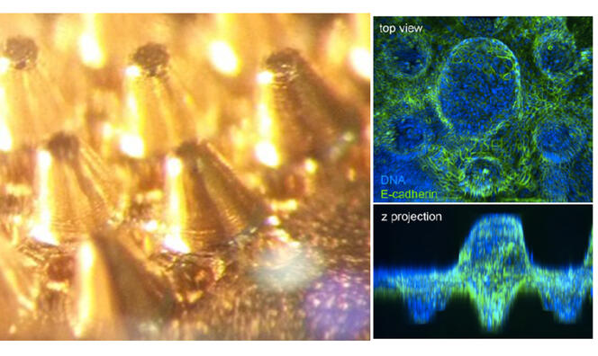
Since moving to Institut Curie in 2011, the scientist and her colleagues have been developing particular organs on a chip, or “tumours from patients on a chip”. These are “micro-tumours created using different cells from the same patient; not only cancer cells but also others that are naturally present in tumours, such as immune cells and blood vessel cells”, she explains.
Using this type of tool, the scientist hopes to fulfil her dream of developing personalised medicine systems that could test a patient’s response to chemotherapies or immunotherapies (two types of cancer treatment), as these may be more or less effective depending on the characteristics (notably genetic) of each tumour. “If we manage to develop these tools, they could mean that patients are directly given the most effective therapy for them, thus increasing their chances of survival,” she hopes.
During a recent study4, Descroix and her colleagues demonstrated the feasibility of this concept in some ten patients. It is now necessary to repeat the study on a larger scale, so a major clinical trial is being planned in around two hundred patients, and should start within the next six months.
Numerous discoveries are anticipated
Applied research is not the only area being addressed. “My team is also carrying out fundamental research to improve our knowledge of organs and their diseases. To be able to work on both sides of research – which tend to be opposed although they feed off each other – is particularly close to my heart,” Descroix points out with pride.
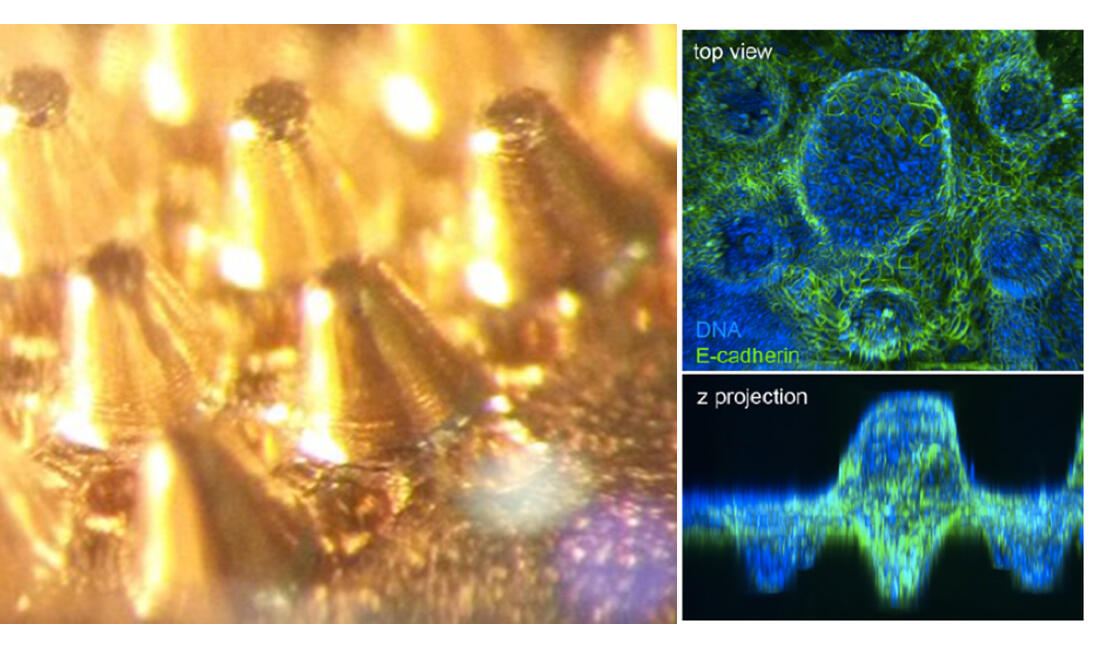
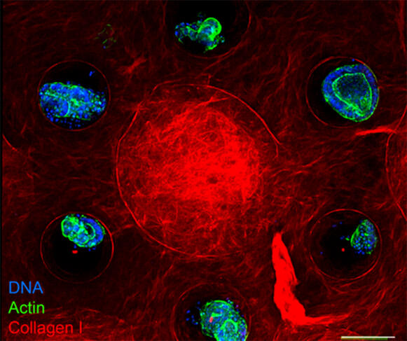
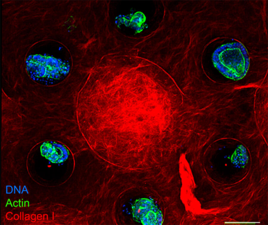
In this area, the scientist is particularly “enjoying” trying to answer some highly specific questions on the intestine, “a superb organ that is too often underestimated”. For example, during recent research work, published notably with Danijela Vignjevic, a cell biologist at Institut Curie5, she co-developed an intestine on a chip that provided information on the development of the different cell types in the intestinal epithelium, which lines the internal surface of the small intestine. “We wanted to know what drives the spatial organisation of these different cell types, in the knowledge that they are not placed randomly but at precise levels in the epithelium,” she adds.
And the scientist and her team hit the jackpot. They were able to demonstrate that if the particular geometry of the epithelium in crypts (grooves) and villi (protrusions) partly regulates the positioning of cells in this tissue, this is not sufficient. It is also necessary for particular cells, or fibroblasts, to be present, as these produce substances (growth factors and collagen, the principal constituent of the extracellular matrix) that are essential to the correct positioning of cells. Indeed, Descroix concludes, “organs on a chip have enormous potential. In the coming years, they should enable a huge number of discoveries, in both fundamental and applied research and in the clinic!” ♦
See on our website
Mini-organs with maximum potential
- 1. Laboratoire Physique de la Cellule et Cancer (CNRS/Institut Curie/Sorbonne Université). “Macromolécules et Microsystèmes en Biologie et en Médecine” team, based at the Institut Pierre Gilles de Gennes Pour la Microfluidique (IPGG) in Paris.
- 2. “Faire mieux avec moins: la microfluidique!”, Stéphanie Descroix, Jean-Baptiste Salmon, Julien Legros, in Étonnante chimie, under the direction of Claire-Marie Pradier, CNRS Éditions, 2021.
- 3. Laboratoire Physico-chimie des Électrolytes, Colloïdes et Sciences Analytiques (CNRS / École Supérieure de Physique et de Chimie Industrielles / Sorbonne Université).
- 4. Irina Veith et al. bioRxiv. 21 June 2023. doi: https://doi.org/10.1101/2023.06.21.545960
- 5. Marine Verhulsel et al. Lab Chip. 27 January 2021. https://pubs.rsc.org/en/content/articlelanding/2021/lc/d0lc00672f
Explore more
Author
A freelance science journalist for ten years, Kheira Bettayeb specializes in the fields of medicine, biology, neuroscience, zoology, astronomy, physics and technology. She writes primarily for prominent national (France) magazines.


