You are here
Facial Recognition: Cracking the Brain’s Code
Even in the middle of a crowd, we manage to identify—instantly and often without even thinking about it—the people we know. In contrast to the ease with which we recognize faces, the underlying cerebral mechanisms are extremely complex and poorly understood. In June of this year, the laboratory headed by Dr. Doris Tsao at the California Institute of Technology achieved a breakthrough in the understanding of these mental processes. Published in the journal Cell, the results of this study1 show that it takes only a few hundred neurons, each one encoding a specific physical characteristic, to recognize a face.
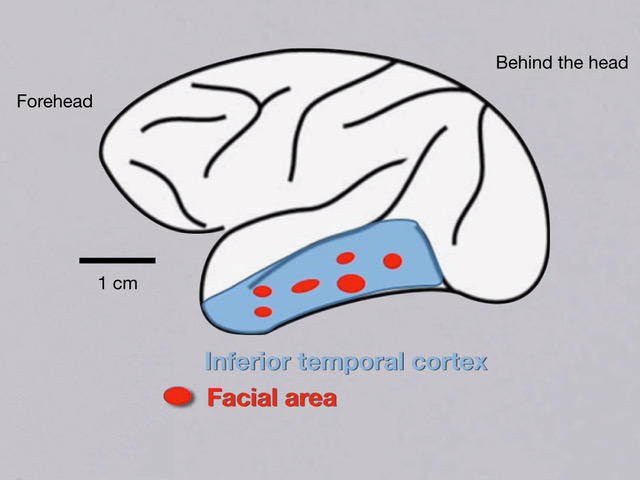
The inferior temporal cortex, the center of facial recognition
Starting from the beginning, which neurons are activated when we look at a face? In a previous study,2 Tsao used the functional MRI (Magnetic Resonance imaging) technique to detect changes in the blood flow of the brain, resulting from an increase in neuronal activity. The authors measured the cerebral activity of monkeys presented with facial images as well as random objects and shapes. Their findings showed that facial identification is concentrated in six small areas of the inferior temporal cortex, called “face patches”. Within these zones, certain neurons, also called "face cells," are strongly activated when a face, rather than any other object, is presented to the subject.

How do face cells react in response to a face?
Until now, the predominant facial recognition model accepted by the scientific community was the “grandmother cell.” According to this theory, every face (e.g. your grandmother’s) is represented by a specific face cell. However, this model has a major flaw: the world’s population now exceeds 7 billion, which is more than the face cells in the inferior temporal cortex! According to this new research by Chang and Tsao, each face cell encodes a particular characteristic, describing the face’s spatial features or other factors related to its appearance.
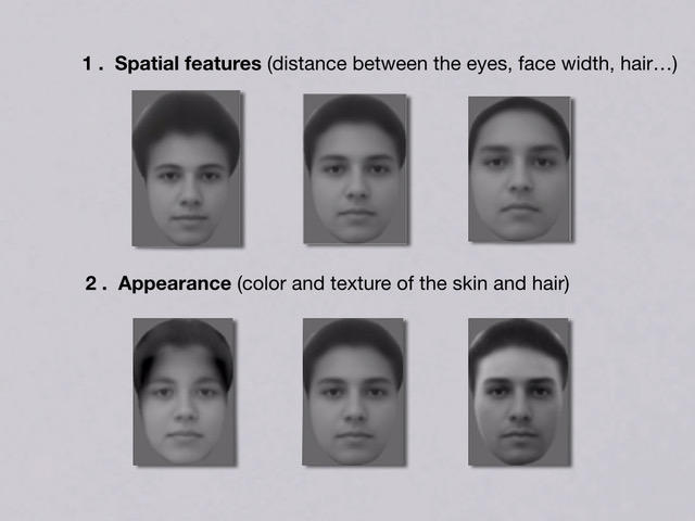
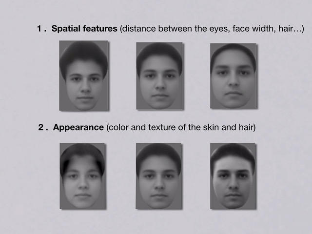
Based on the analysis of a database of 200 faces, the researchers began by identifying 50 parameters that vary the most from one face to another, including 25 representing its spatial features (face width, distance between the eyes…), and 25 corresponding to its appearance (i.e. the color and texture of the skin and hair). These 50 characteristics can be associated to reproduce any face, similar to the way that red, blue and green tones can be combined in different proportions to create any color.
Among all the possibilities created from these 50 parameters, 2000 faces were selected at random to be presented to two monkeys (rhesus macaques), while the researchers recorded the activity of the face cells in three zones, using very fine electrodes implanted in the inferior temporal cortex. A total of 205 neurons were monitored. By analyzing their activity, the authors were able to show that each face cell responds to a specific characteristic. For example, if a cell responds when the eyes are widely spaced, it is activated each time this feature is detected, even when the two faces look very different overall. In addition, certain face patches are more sensitive to the appearance of the face, while others are more easily activated by its spatial features.
It only takes about 200 neurons to identify any face
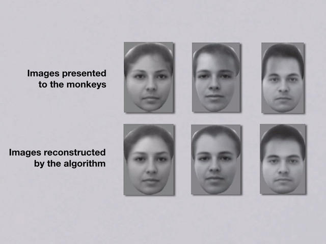
To test the robustness of this code, Chang and Tsao then reversed the process, seeking to predict the appearance of faces seen by the monkeys, based on the activity of their face cells. To do this, they fed an algorithm with the activity of the 205 cells that had been monitored during the presentation of faces from the database. The monkeys were then shown features that they had never seen before. Quite surprisingly, the predictions made using the algorithm looked almost exactly the same as the actual faces.

It would therefore seem that it takes only 205 neurons to encode any given face—a very small number considering the complexity of the task! It is, of course, highly probable that other neurons are involved in representing certain subtler characteristics, but most of the work seems to be performed by these 205 cells. This tells us that the brain has adopted an extremely effective facial recognition code, both simple and sophisticated, involving a minimum of neurons but enabling the recognition of any face.
Why is it so important to crack this code?
This breakthrough opens new perspectives for exploring the functions of other cerebral areas that are associated with object recognition, as well as non-visual sensory systems such as taste and touch. From a clinical point of view, these results could shed light on the origin of prosopagnosia, a pathology whose sufferers find it difficult or even impossible to identify or memorize human faces.
In addition, this code could have applications in forensic medicine, for example in the production of facial composites. It is extremely difficult for a witness to describe a face precisely in words, down to its smallest details—especially when in a state of shock. Now it might be possible to reconstruct a face by asking the witness to visualize the criminal and analyzing the resulting cell activity. However, although the researchers believe that facial perception is similar in monkeys and humans, the same kind of study would be inconceivable in humans due to its highly invasive nature. Few if any test volunteers would agree to having 205 electrodes implanted in their brain…
The analysis, views and opinions expressed in this section are those of the authors and do not necessarily reflect the position or policies of the CNRS.


