You are here
Preventing malaria from proliferating
03.08.2024, by
Malaria affects more than 247 million people throughout the world and may have caused up to 620,000 deaths in 2021. Under the ROAdMAP project¹, Ana Gomes and her team at the Laboratory of Pathogens and Host Immunity² in Montpellier (southern France) are seeking to understand how the parasite responsible for this disease replicates its DNA, in a bid to discover any weaknesses that could be exploited as new therapeutic strategies.
1. DNA replication initiation in malaria parasites.
2. LPHI (CNRS / Université de Montpellier).
1
Slideshow mode
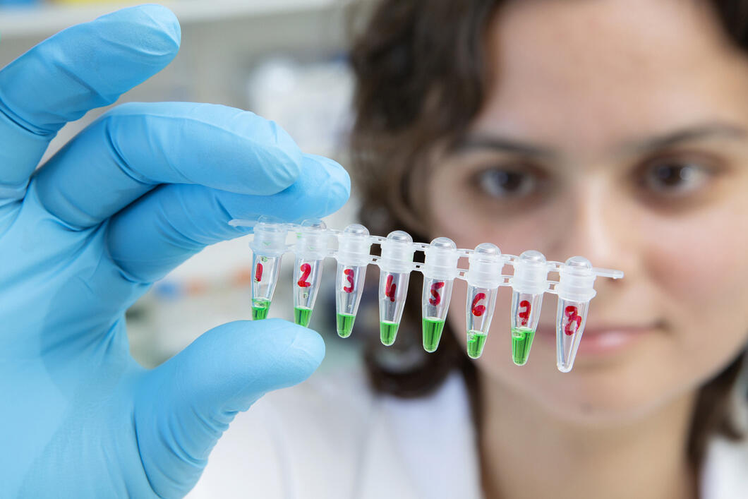
"Plasmodium falciparum", the parasite that causes malaria, proliferates by producing dozens of daughter cells during a single cell cycle, unlike yeasts or human cells, for example. During this process, each parasite replicates its genetic material several times thanks to the specific genes being studied by the scientists. To achieve this, the team is using molecular cloning to produce DNA that contains non-functional copies of these genes.
Christophe Hargoues / LPHI / CNRS Images
2
Slideshow mode
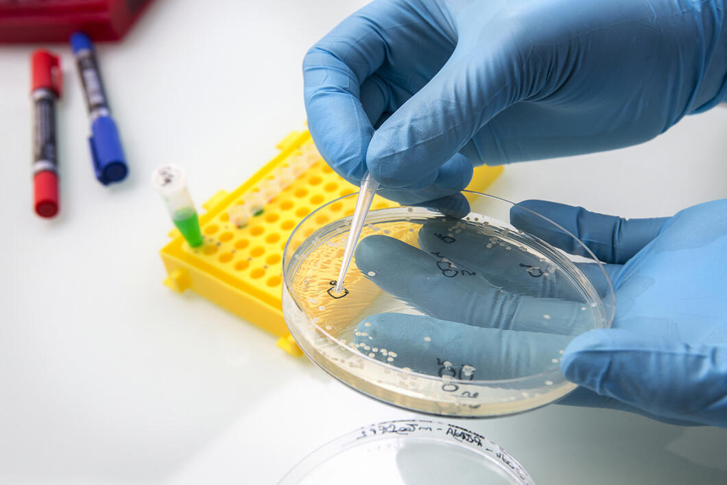
Once the mutated DNA has been reproduced, the scientists use bacteria to amplify and obtain it in large quantity. To do so, they introduce the DNA into bacteria applied to plates coated with agar, a gelling agent that allows them to develop. The researchers can then determine which bacteria have succeeded in producing the mutated DNA of the parasite.
Christophe Hargoues / LPHI / CNRS Images
3
Slideshow mode
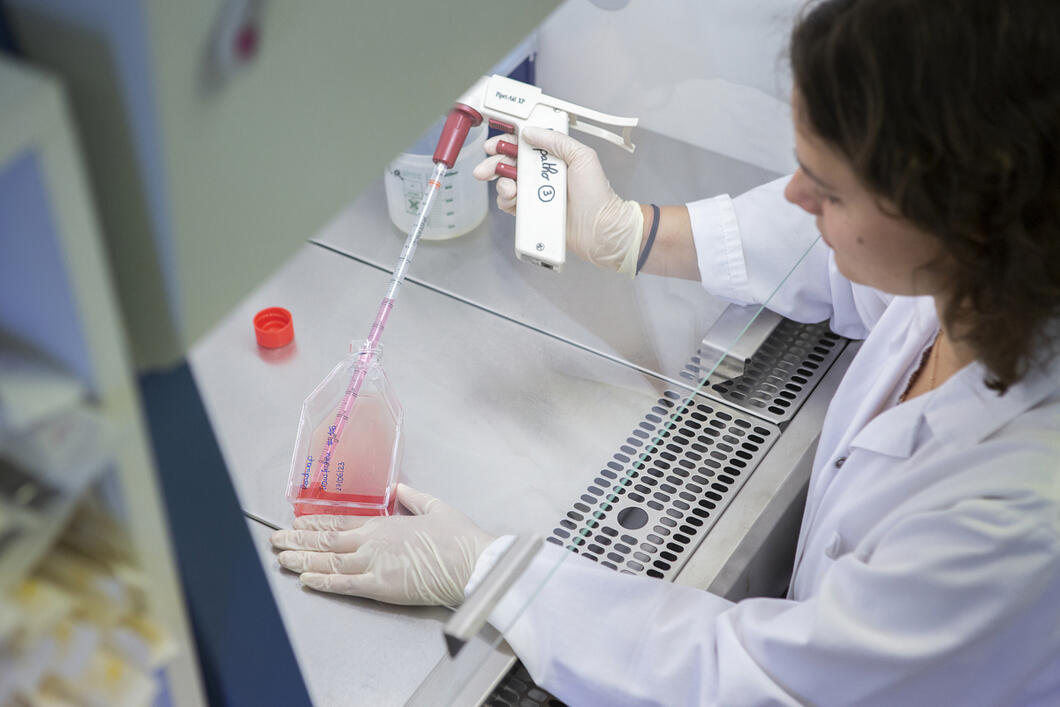
The scientists cultivate "Plasmodium falciparum" in vitro: it develops within human red blood cells in a rich culture medium that is similar to the blood environment. Each day, this culture medium (seen here) needs to be replaced to provide new nutrients. All manipulations are carried out under sterile conditions to prevent contamination by bacteria or other micro-organisms.
Christophe Hargoues / LPHI / CNRS Images
4
Slideshow mode
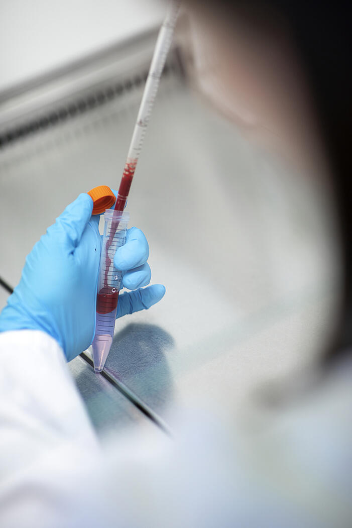
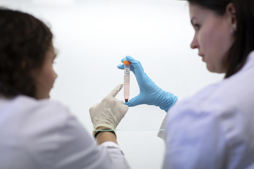
To study specific stages in the development of the parasite, the scientists are focusing on variations in density between red blood cells infected by different forms of the pathogen. For example, those containing mature forms of "Plasmodium falciparum" are less dense than those containing younger forms. Infected red blood cells are therefore placed on a density gradient, a technique that enables their precise sorting.
Christophe Hargoues / LPHI / CNRS Images
5
Slideshow mode
Once the red blood cells have been placed on the density gradient, and following a centrifugation step, the mature (less dense) cells will gather within a precise zone of the gradient.
Christophe Hargoues / LPHI / CNRS Images
6
Slideshow mode
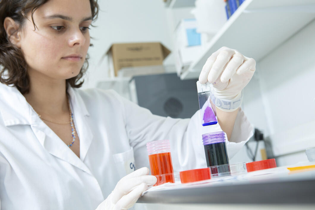
To monitor the density and developmental stage of the parasites, the scientists prepare a blood smear on a glass slide. This adds contrast and improves visualisation of the pathogens within the red blood cells. Staining is a two-stage process: the smear is first dipped in a solution of eosin (a pink stain) that enables the researchers to better see certain parts of the parasite, and then in a solution of methylene blue, which stains its DNA violet. This also makes it possible to count the pathogens and determine their age thanks to the number of nuclei containing DNA.
Christophe Hargoues / LPHI / CNRS Images
7
Slideshow mode
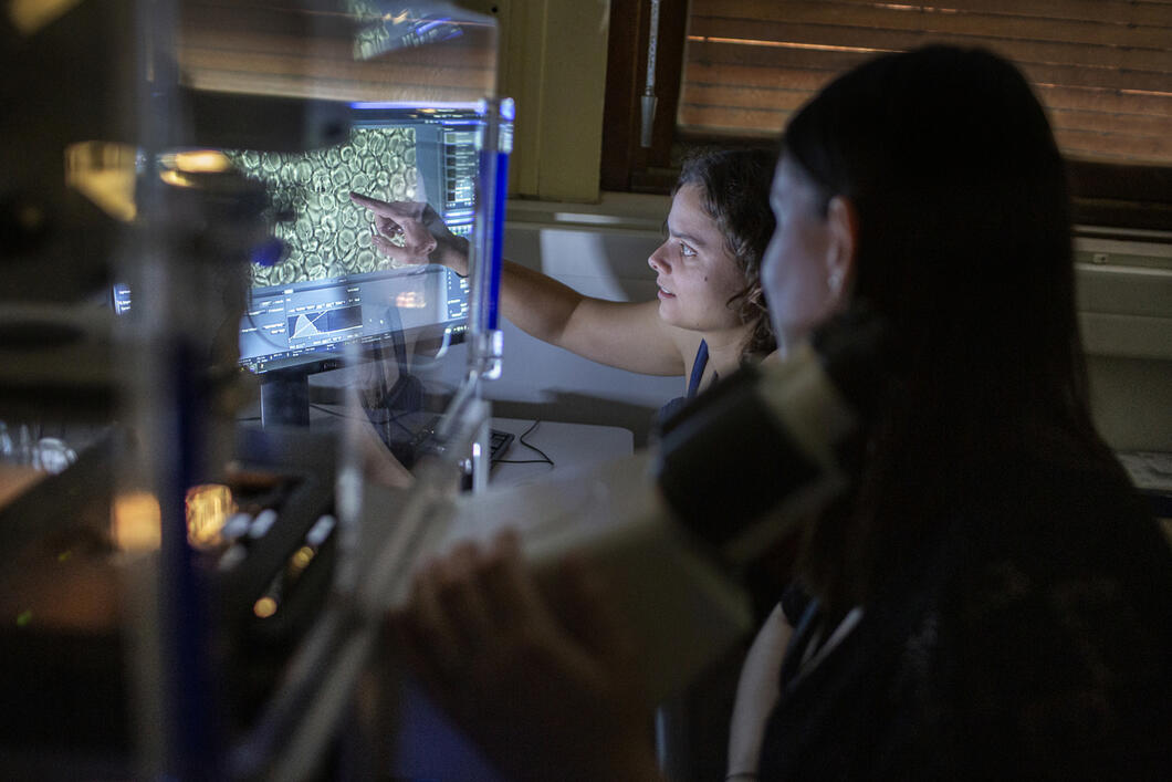
The scientists use a fluorescence microscope to follow the development of "Plasmodium falciparum". The stained proteins enable them to determine the different stages of growth by observing where these colours appear in the cellular structure of the parasite.
Christophe Hargoues / LPHI / CNRS Images
8
Slideshow mode
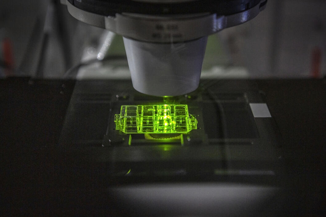
A high-resolution microscope is used to understand how the parasites develop. These are placed in a well on the slide and subjected to a laser that excites a fluorescent protein they contain. The latter absorbs the laser light and in return transmits a light source that renders it visible. The results are then analysed in order to elucidate the DNA replication rate during the different developmental stages of the parasite. The aim is to eventually be able to identify targets for future treatments and contribute to the decline of malaria across the world.
Christophe Hargoues / LPHI / CNRS Images
À propos
Explore more
Life
Article
01/28/2026
Article
11/26/2025
Article
11/24/2025
Article
09/24/2025
Article
09/01/2025
Genetics
Article
01/12/2022
Article
06/19/2017


