You are here
Landscapes of the microworld
08.12.2021, by
Progress in microscopy continues to expand our window onto the world of the infinitesimally small. Whether in chemistry, biology, engineering or digital simulation, these images paint a phantasmagoric and yet factual portrait of the world that we live in, that we build, and that lives within us.

1
Slideshow mode
A raging cubist sea surges forth during the recrystallisation of sodium thiosulfate, a chemical compound used in the mining and chemicals industries, and as a medicinal drug. This image was produced using analysed polarised light.
A. Jeanne-Michaud/IMPMC/CNRS Photothèque
2
Slideshow mode
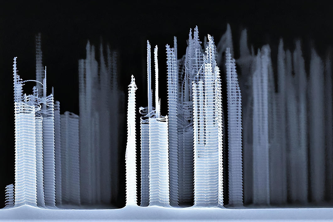
A ghostly cityscape emerges on a silicon sample subjected to plasma etching. In this microelectronic experiment, the presence of a contaminant in the material resulted in a manufacturing defect, creating grooves that resemble the floors of skyscrapers.
Pierre Albert/LN2/IMS

3
Slideshow mode
The relentless war between human macrophages and HIV is observed under a fluorescence microscope. The data thus gathered is used to analyse the strategies deployed by the attackers (in red). In this case, the immune defence forces (blue and green) have also been infected by the agent that causes tuberculosis.
Shanti Souriant/IPBS/CNRS Photothèque
4
Slideshow mode
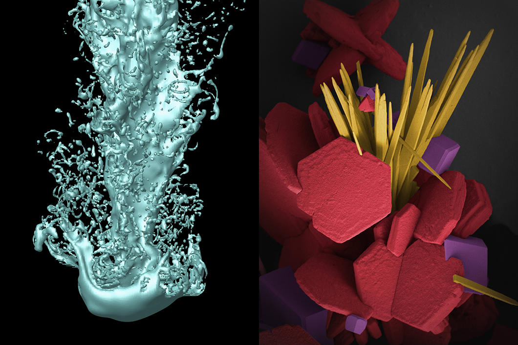
Impossible to capture in situ, this image of a spray of liquid (left) was created with the help of a supercomputer to reproduce the conditions at the nozzle of a diesel fuel injector. In contrast, the solid, static agglomerations of calcium carbonate crystals (right) can be observed directly with a scanning electron microscope. False colours were added to differentiate the crystals.
Alain Berlemont/Sébastien Tanguy/Thibaut Ménart/CORIA/CNRS Photothèque - Bertrand Rebière/ICGM/CNRS Photothèque
5
Slideshow mode
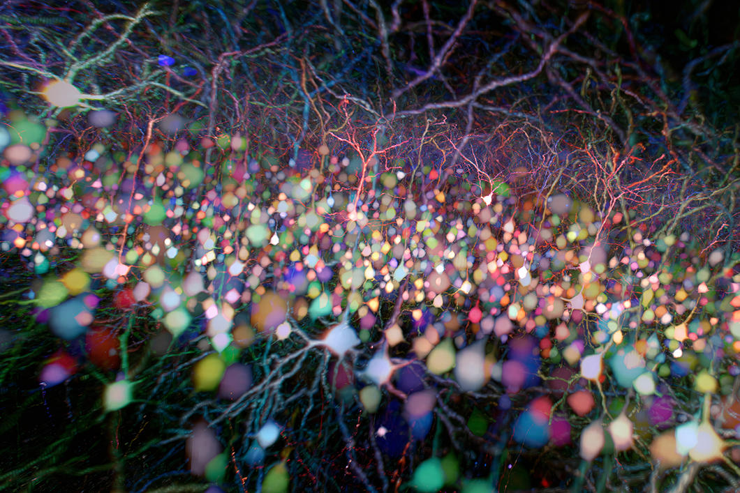
This profusion of fibrous “drops” comes from the brain of a type of transgenic mouse called a “Brainbow”. Interesting phenomenon: the nerve cells seen here under a multicolour multiphoton microscope themselves produce hundreds of fluorescent molecules of different colours, enabling researchers to study the neural circuits in action.
Lamiae Abdeladim/LOB/Institut de la vision/CNRS Photothèque
6
Slideshow mode
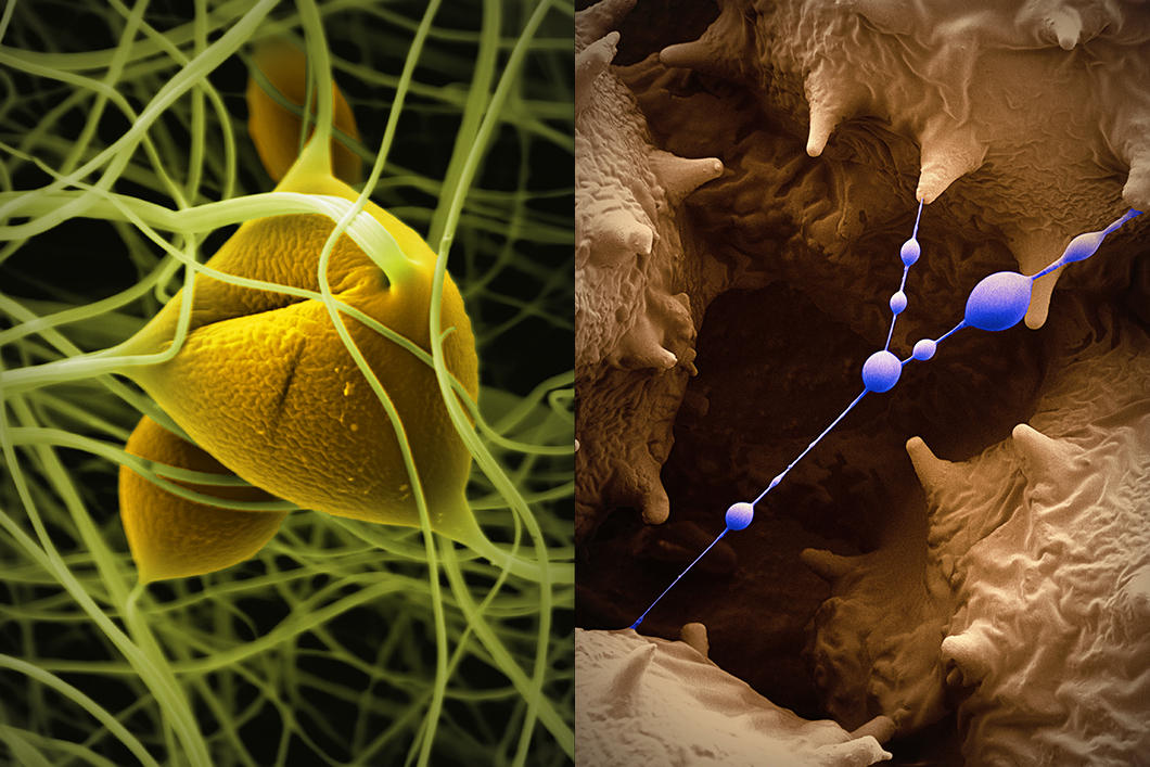
A jungle of vines grows on microscopic scale. On the left are polyacrylonitrile fibres created in the laboratory to be used in the manufacture of biofuel cell electrodes. The fibres on the right were produced in a spider’s abdomen to hold three pollen grains in place. Since scanning electron microscopes only produce black and white images, the colours have been added.
Bertrand Rebière/ICGM/CNRS Photothèque

7
Slideshow mode
Under splashes of copper, a bit of iron. We are looking at the surface of a two-centime coin whose composition is being analysed using LIBS (laser-induced breakdown spectroscopy): laser pulses produce a spark on the surface that reveals the chemical profile of the material. The technique is used, for example, to determine the chemical composition of prehistoric paints, hazardous materials and Martian rocks.
Alain Queffelec/Léna Bassel/Pacea/CNRS Photothèque

8
Slideshow mode
This tangle of pop art tubes comes from… a fruit fly’s intestines. The insect’s digestive system has been coloured blue and red and its sex organs (in this case male) in green and yellow. Obtained with a confocal microscope, the image can be used to study the complex and surprising links between the two biological systems, indicating that the difference between the sexes is also (literally) visceral.
Bruno Hudry/Institut de biologie Valrose/CNRS Photothèque
Explore more
Life
Slideshow
02/25/2026
Article
01/28/2026
Article
11/26/2025
Article
11/24/2025
Article
09/24/2025
Biology
Article
09/24/2025
Article
05/25/2025
Article
03/26/2025
Article
12/02/2024
Article
10/15/2024
Microscopy
Article
05/23/2024
Slideshow
11/10/2022


